Pediculectomy and left-sided hemilaminotomy in a canine with Hansen type I disc herniation between T13-L1 intervertebral levels
Main Article Content
Abstract
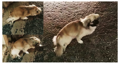
Wiski, a male mongrel dog (Pekingese breed) of four-year-old male, arrived for consultation. He presented paraplegia and signs of acute spinal pain. The initial protocol was to treat the emergency with fluid therapy, pain management and blood pressure control. Neurological examination revealed upper motor neuron signs in the pelvic limbs, compromised proprioception and urinary function, severe pain at the thoracolumbar junction with a reduced panniculus reflex. Imaging studies (X-ray and CT) confirmed a Hansen type I disc herniation between the thirteenth thoracic vertebra and the first lumbar vertebra. A conventional approach for hemilaminectomy was carried out between T13 and L1 intervertebral levels. A first exploratory pediculectomy was performed on the left side of L1 vertebral pedicle, which revealed disc material in the cranial portion; hence a second pediculectomy was started at T13. Both pediculectomies were joined by hemilaminectomy. Disc material was removed from the vertebral canal and washed with saline solution to finish the synthesis of the muscular planes, thoracolumbar fasciae, subcutaneous tissue, and skin. Postoperative period involved total restriction of movement, antibiotherapy and anti-inflammatory and analgesic therapy. Urinary retention, associated to upper motor neuron signs, was treated with Diazepam exclusively during the first days. During the first 14 days, the animal showed no evidence of pain, sepsis or surgical complications. At 23 postoperative days, the animal recovered the motor activity of its pelvic limbs.
Article Details

This work is licensed under a Creative Commons Attribution-NonCommercial 4.0 International License.
National Center for Animal and Plant Health (CENSA)