Clinical epidemiological study of the retained ulnar cartilage cores in canines from Havana
Article Sidebar
Main Article Content
Abstract
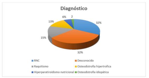
This study was aimed at calculating the frequency of the retained ulnar cartilage cores and determining risk factors and percentage of clinical and radiological signs present in canines treated in a veterinary assistance service in Havana, from January 2017 to March 2020. The sample was composed of canines from three to six months of age with a radiological diagnosis of retained ulnar cartilage cores (RCC). The influence of breed, sex and age on the diagnostic outcome was determined by Chi-square test. In addition, RCC frequency was calculated based on radiological signs and findings, and RCC percentage was determined according to feeding and calcium supplementation. Chi-square test showed association between disease and breed (p≤0.05). The most frequent signs were the presence of cartilage on X-rays (100 %), radial anteversion (97.06 %), pain (91.18 %), ulnar shortening (88.24 %), ulnar styloid process dislocation (82,35 %), claudication (67.65 %), mild (58.82 %) and moderate (23.53%) carpal valgus, supination (50 %), elbow incongruity (41.18 %), osteoarthritis (8.82 %), and hyperextension (11.76 %). RCC frequency of presentation was high reaching values of 32.38 %. The most frequent signs were the presence of cartilage on X-rays, anteversion, pain, ulnar shortening and proximal dislocation of the styloid process. Furthermore, 35.29 % of the animals were overfed and 70.59 % were supplemented with high-dose calcium.
Article Details

This work is licensed under a Creative Commons Attribution-NonCommercial 4.0 International License.
National Center for Animal and Plant Health (CENSA)References
Rueda J, Fernández A. Osteodistrofias en el perro y en el gato, diagnóstico diferencial. AVEPA. 1989; 9 (1): 9. Available from: https://core.ac.uk/download/pdf/33159962.pdf.
Palavecino JC. Retención de cartílago de crecimiento; importancia del diagnóstico y tratamiento precoz. Tandil, Argentina: Facultad de Ciencias Veterinarias UNCPBA; 2018. Available from: www.ridaa.unicen.edu.ar/xmlui/handle/123456789/1593.
Lappalainen A, Pulkkinen H, Mölsä S, Junnila J, Hyytiäinen H, Laitinen-Vapaavuori O. Breed-typical front limb angular deformity is associated with clinical findings in three chondrodysplastic dog breeds. Front. Vet. Sci., 17 January 2023. Available from: https://www.frontiersin.org/journals/veterinaryscience/articles/10.3389/fvets.2022.1099903/full.
Fernández T, Gómez L, Ríos A. Deformaciones angulares en los miembros anteriores del perro como consecuencia de alteraciones del crecimiento del cúbito. Clínica veterinaria de pequeñas especies. 1990; 10 (3): 116-150. Available from: https://ddd.uab.cat/record/69869.
Koch D. Orthopedic problems in growing dog. 2020. Available from: https://dkoch.ch/fileadmin/user_upload/Selected%20handouts%20from%20orthopedic%20surgery/Orthopedic_problems_in_growing_dogs.pdf.
Schaible L. Angular Limb Deformities in Dogs: Types, Causes & Treatment. Hill’s pets nutrition. 18 August 2021. Available from: https://www.hillspet.com/dog-care/healthcare/angular-limb-deformities-in-dogs?lightboxfired=true.
Arias I. Ostectomia en cuña para el manejo quirúrgico de la deformidad angular radio-ulnar distal. Chile: ICBM Facultad de medicina, Universidad de Chile; 2012. pp. 14-27. Available from: https://core.ac.uk/download/pdf/46531543.pdf.
Arnaiz V. Osteodistrofias en perros una visión nutricional. Lima, Perú: Universidad Nacional Agraria La Molina; 2010. Available from: http://tecnovet.uchile.cl/index.php/RT/article/download/15934/16421/0.
Catalano MR. Enfermedad del crecimiento de los perros: el radio curvo [en línea]. UNICEN. Universidad Nacional del Centro de la Provincia de Buenos Aires; 29 April 2014. Available from: http://www.unicen.edu.ar/node/12157.
Kwon M, Kwon D, Lee J, Lee K, Yoon H. Evaluation of the Radial Procurvatum Using the Center of Rotation of Angulation Methodology in Chondrodystrophic Dogs. Front Vet Sci (3):8. 2022. Available from: https://pubmed.ncbi.nlm.nih.gov/35047584/.
Dos Santos C, Lopez A, Texeria L, Garcia D, Matos V, Ribeiro C. Clinical comparison between two stabilization methods in distal tibial angular deviation corrected by the CORA method. Acta de ciencias veterinaria (48):12. 2020. DOI: http://doi.org/10.22456/1679-9216.99560 https://www.researchgate.net/publication/339700018_Clinical_comparison_between_two_stabilization_methods_in_distal_tibial_angular_deviation_corrected_by_the_CORA_method.
Griffon D. Cubital osteotomy and ostectomy. John Wiley & Sons, Inc. January 2016 [cited 01 March 2020]. Disponible en: http://doi.org/10.1002/9781119421344.ch122.
Fossum TW. Small animal surgery. St Luis Missouri, USA: 5ta ED. Elsevier Mosby; 2019.
Demko J, Mclaughlin R. Developmental Orthopedic Disease. Veterinary Clinics of North America Small Animal. 2012; 35 (1): 1111-11. http://doi.org/10.1016/j.cvsm.2005.05.002
Harfush LA. Deformidades angulares en los miembros inferiores. medigraphic Artemisa. 2011; 3 (2): 90-97. https://www.medigraphic.com/pdfs/orthotips/ot-2007/ot072e.pdf
Bottcher P, Brauer S, Werner H. Estimation of joint incongruence in displastic canine elbows before and after dynamic proximal ulnar ostectomy. Vet Surg. 2013; 42 (4): 371-376. http://doi.org/10.1111/j.1532-950X.2013.01085.x
Kaempf R, Ribak S, Farina JP, Aita M, Delgado P. Deformidad de Madelung. Opciones de diagnostico y tratamiento. Rev Iberam Cir Mano 2021;49(2):e140–e154. https://www.thieme-connect.com/products/ejournals/pdf/10.1055/s-0041-1739452.pdf?articleLanguage=es
Silva-molano RF. Osteocondromatosis en caninos, descripción de un caso clínico. vet.zootec. 2010; 4 (2): 9-14. http://vip.ucaldas.edu.co/vetzootec/downloads/v4n2a01.pdf
Benjamino K. Juvenile Canine Orthopedic Diseases: Part 1: thoracic limbs. PVMA Spring Clinic. 2018; 12 (1): 13-20. https://cdn.ymaws.com/www.pavma.org/resource/resmgr/docs/Spring_Clinic_Proceeding/Benjamino/Juvenile_Canine_Orthopedic_D.pdf
Villaluenga JE, Godinho P, Puchol JL. Ostectomía dinámica distal de cúbito (Distal Dynamic Ulnar Ostectomy o DDUO) Trabajo de revisión. 2021. https://axoncomunicacion.net/wp-content/uploads/2021/10/cv-101.pdf
Engdahl C, Houglund O, Hedhammar A, Hanson J. The epidemiology of osteochondrosis in an insured Swedish dog population. Preventive Veterinary Medicine Volume 228, July 2024. https://doi.org/10.1016/j.prevetmed.2024.106229
Yuan E, Jamarillo D. Imaging of osteochondrosis. Pediatric radiology. 2019; 49 (12):1610-1616. http://doi.org/10.1007/s00247-019-04556-5
Simón F, López F, Teixeira L, Aparecida L, Lourenço L, Garcia DM, Saldanha D, Barbosa EO. Treatment of Radius Curvus in a Young Dog with Association of Radial Physeal Stapling, Ulnar Ostectomy and Transarticular Dynamic External Fixator CASE REPORTS (SUPPLEMENT) (49): july, 2021. https://doi.org/10.22456/1679-9216.105684
Vezzoni A. Osteotomies for treating Young dogs with incongruent elbow joints. Asociacion britanica de veterinarios de pequeños animales. 2015. pp. 196-197. http://doi.org/10.22233/9781910443521.19.1
Harari J. Osteopatias del desarrollo en perros y gatos. Manual de MSD. 2020. https://www.msdvetmanual.com/es/sistema-musculoesquel%C3%A9tico/osteopat%C3%ADas-en-peque%C3%B1os-animales/osteopat%C3%ADas-del-desarrollo-en-perros-y-gatos